Glass
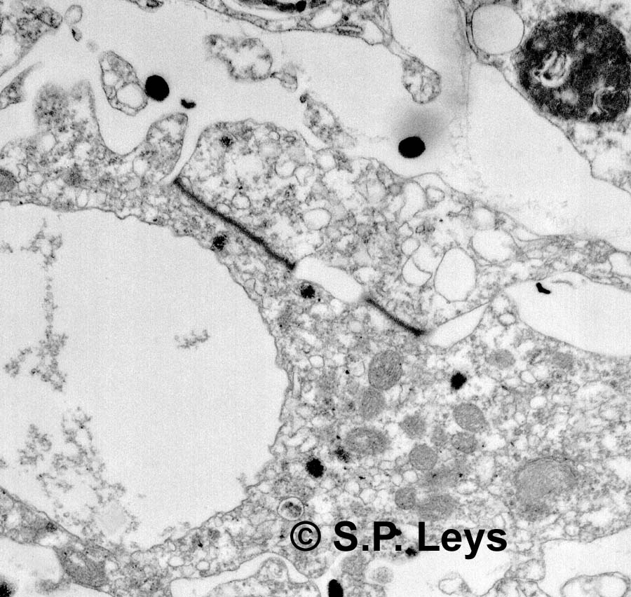
sponges begin as cellular embryos, like other metazoans, but after the
5th cleavage cells fuse, so that eventually a giant multinucleated cell
is formed. There are some regions (rightly called cells) that have single
nuclei, but these are always connected to the multinucleated tissue
by open or plugged cytoplasmic bridges. The plugs are an odd osmiophillic
junction, called a ‘plugged junction’ (dark band seen in electron micrograph
at right). |
 |
|
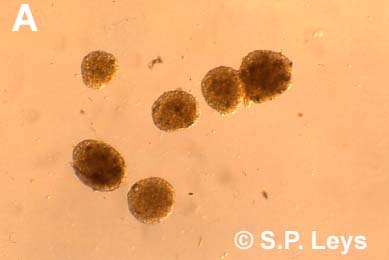
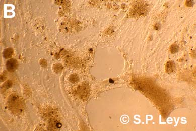
| To understand how the tissue can be made up of a giant cell, the tissue can be dissociated through fine mesh (Nitex for example) and allowed to come back together. Similar experiments with cellular sponges provided the first illustration of cell adhesion molecules. When tissue from the glass sponge Rhabdocalyptus dawsoni (the boot sponge - right) is dissociated it forms opaque aggregates (A, below) that are syncytial. If the dissociated tissue is cultured on an extract made from the sponge itself, it adheres, and spreads to form a giant, single thin tissue (B, below). |  |
|
  |
||
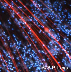
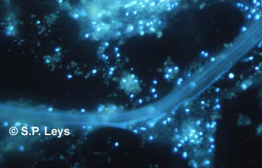
| Labelling the nuclei with a fluorescent stain (blue - Hoechst) and the microtubules with an anti-body to tubulin (red) shows that the syncytial tissues are extensive in adherent cultures. Nuclei (blue) in living preparations move through the tissue on vast bundles of microtubules and leave a trail of light marking their path | ||
 |
 |
|
| Nuclei and microtubule cytoskeleton | Nuclei
moving in cytoplasmic streams |
|
| From Leys, 1999, Biol. Bull. 188:241-254 | ||